Labeled Dental X Ray Machine Parts

Requires a way to obtain the size and form of craniofacial structures in the patient.
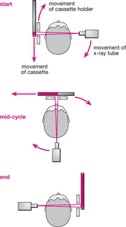
Labeled dental x ray machine parts. A standard 2d panorex will provide all of the imaging requirements needed for such treatments as caries detection diagnosis of tmj issues opg images and images of the patients entire detention in a dental x ray. X ray has three main components. Which dental x ray machine is right for me. X rays are also used for treatment of tumours and abnormal condition in the body.
A 1954 b 1964 c 1974 d 1984. Dental radiographs are commonly called x rays. A flat disk with a small opening that restricts the size of the x ray beam emitted maximum diameter of opening by law is 2 75 cm. Other sets by this creator.
Quality affordable used recycled dental x ray equipment can help you with your new or expanding practice. To determine if dental x ray film is fresh process the film using fresh chemicals. C 1974 2 dental receptors placed inside the mouth are termed. The uses of x ray in industries are as.
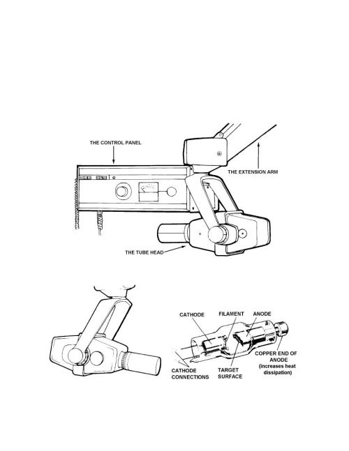
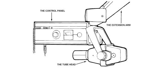
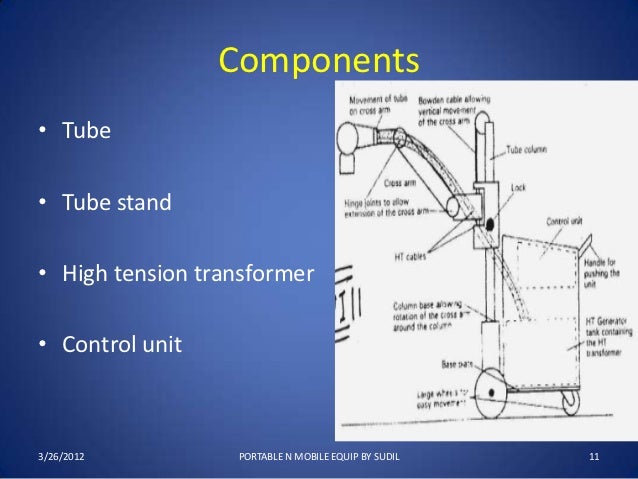
Dentists use radiographs for many reasons. Syed mustafa jamal 2. Suspends the x ray tubehead and. We can even install your purchased equipment if you need assistance.
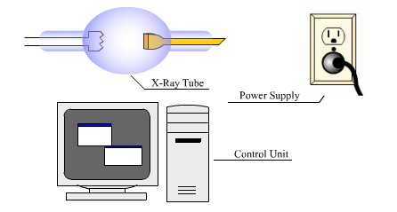
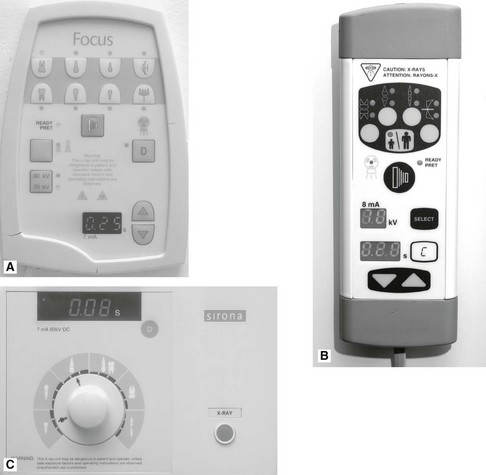
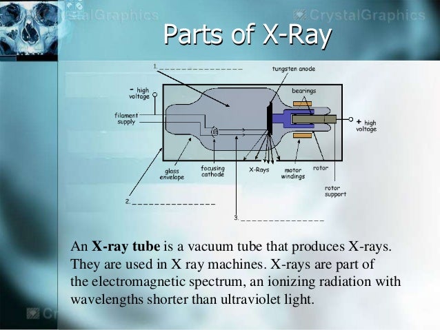
Parts and definitions of a dental x ray tube learn with flashcards games and more for free. X rays are no doubt used extensively in modern medicine for detection of fracture in bones presence of tumour infection of lungs kidneys and other organs of body. All of the above. Operating console high frequency generator x ray tube internal external other parts include collimator and grid bucky x ray film 3.
The positive electrode in the x ray extension arm a part of the dental x ray machine. X ray machine was invented by henry backral of germany. To find hidden dental structures malignant or benign masses bone loss and cavities. Dental planet has the x ray machine parts and x ray machine accessories to keep your practice running smoothly all at the lowest prices anywhere.
T f after exposure each mount should be labeled with the patient s name and the date. Components of x ray machine 1. A radiographic image is formed by a controlled burst of x ray radiation which penetrates oral structures at different levels depending on varying anatomical densities before striking the film or sensor. Dental x ray tube head diagram.
We usually have items such as recycled wall mount equipment pano equipment intraoral sensors and cassettes. A intraoral b extraoral c occlusal d all of the above. Anatomy of the x ray machine.


